ODDZIAŁY
Description of the service
Dental treatment at enel-med is always preceded by detailed diagnostics. We perform the diagnostics using the latest methods and equipment. Depending on your needs, this includes the following:
Orthopantomogram
Orthopantomogram is an X-ray scan that is the basis for dental diagnostics, planning, and control of treatment results. It shows most of the structures within the maxilla and the mandible, and ensures the dentist sees the areas that cannot be visually inspected during periodic dental check-ups.
Orthopantomogram is used, among others, in endodontics and dental surgery.
Lateral cephalometric radiograph
Lateral cephalometric radiograph is a radiograph that shows the lateral proportions of the face. It is indispensable in planning the orthodontic treatment; it enables measurements that will be used to select the right braces, and helps estimate the time necessary for successful treatment of malocclusion.
Periapical X-ray
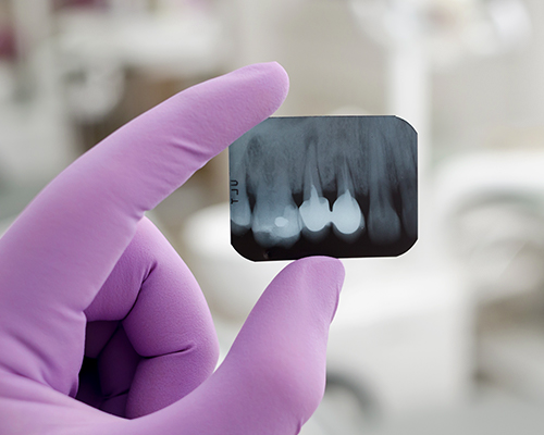
Periapical X-ray is a scan of individual teeth and surrounding tissues. It accurately shows caries present in the teeth and the inflammation around the root apices. Due to the high resolution, the scan is very accurate, showing the number and course of canals and roots, invisible during a routine dental check-up.
Periapical X-ray is used in diagnostics and root-canal treatment.
T-scan
T-scan is a digital occlusal analysis system, recording the patient’s occlusal condition and analysing the chewing and biting forces in the oral cavity. All the measurements are displayed on the computer screen, making it easy to see the patient’s situation. t-scan
T-scan is often used by prosthodontists and orthodontists because it detects occlusal overloads that might cause pain, and enables much more accurate treatment.
Facial arch
The facial arch accurately records the proportions and relations of the upper and lower teeth with respect to other anatomical elements of the skull, e.g. the temporomandibular joints. Measurements taken with the facial arch are transferred to plaster models created in a lab.
The examination is time-consuming (30 to 60 minutes), but painless. It is the basis for planning and performing prosthetic or implant-prosthetic treatment.
Impressions
Impressions recreate the patient’s teeth using a special mass applied in a mould (an impression spoon). They provide information about conditions present in the patient’s oral cavity, and are easy to understand by a dental technician who performs the prosthetic work.
Impressions are used in various fields of dentistry. Orthodontists use them to create plaster models necessary for diagnostics and treatment planning. The prosthodontist needs them to make dentures, (permanent and temporary) crowns, bridges or dental guards. An impression is also ordered before preparing teeth whitening trays.
Photos
Photos are taken at each stage of the treatment to record it, and also to observe and compare obtained results. Profile, side and smiling photos enable to analyse the facial features, whereas internal photos show the relations of all the teeth and the selected areas. By examining photos on the screen in high magnification, the patient becomes more aware of their health and aesthetic needs.
Photos are used in every field of dentistry, most commonly to compare the condition before and after whitening, placing veneers, professional cleaning, curettage, orthodontic treatment, extensive and bone regeneration.
Dental microscope
The dental microscope allows for multiple magnification of the examined tooth surface and oral mucosa. The obtained image significantly increases the effectiveness of treatment, also making it easier to carry out the right diagnostics regarding the presence of caries, without having to take X-ray scans.
The microscope is used in the fields that particularly require precision, e.g. root-canal treatment and periodontics.
When the doctor needs a broader field of vision at a lower magnification, dental loupes (either worn on the forehead or integrated with glasses) can replace the microscope. Loupes significantly reduce the strain on the eyes, and are readily used by prosthodontists (in order to remove a portion of a tooth for aesthetic prosthetic work), surgeons, periodontists (for bone reconstruction procedures) and even by hygienists when cleaning teeth.
Dental loupes
Dentists use loupes when they need a broader field of vision at a lower magnification. Used with a forehead band or integrated with glasses, loupes reduce the strain on the eyes, and serve a similar function as a dental microscope. They are used by prosthodontists (when they need to remove a portion of a tooth for aesthetic prosthetic work), periodontal surgeons (for bone reconstruction procedures) and hygienists when cleaning teeth.
Contact form
Please complete the form below. We will call you back, tell you about the details of the offer and arrange an appointment for you.


